Pleural Mesothelioma Electron Microscopy / Mesothelioma Spheroid Sem Stock Image C030 3472 Science Photo Library : In a previous publication the ultrastructure of pleural effusions in cases of pleural mesothelioma was reported.
Pleural Mesothelioma Electron Microscopy / Mesothelioma Spheroid Sem Stock Image C030 3472 Science Photo Library : In a previous publication the ultrastructure of pleural effusions in cases of pleural mesothelioma was reported.. Asbestos exposure of 131 patients with pleural malignant mesothelioma in the paris area to. (b,c) tnts connecting primary malignant cells . The epithelioid, sarcomatoid and biphasic types (8). Furthermore, p16 deletions were specifically detected by fluorescence in situ hybridization, and electron microscopy showed numerous, . Histologically, three types of malignant pleural mesothelioma (mpm) are classically recognized:
(b,c) tnts connecting primary malignant cells . Risk estimates and whither electron microscopy for diagnosis? The same method has now been applied to a . Asbestos exposure of 131 patients with pleural malignant mesothelioma in the paris area to. (a) scanning electron micrograph of two separate mesothelioma cells tethered by a nanotube.
Pleural Mesothelioma Clouded Leopard Note The Postiive Download Scientific Diagram from www.researchgate.net The results of a light and electron microscopic study and enzyme histochemistry of reactive mesothelial cells and diffuse and localized (solitary) pleural. The same method has now been applied to a . (a) scanning electron micrograph of two separate mesothelioma cells tethered by a nanotube. Furthermore, p16 deletions were specifically detected by fluorescence in situ hybridization, and electron microscopy showed numerous, . Analysis of lung tissue by electron microscopy. (b,c) tnts connecting primary malignant cells . The epithelioid, sarcomatoid and biphasic types (8). No electron microscopy was done on the original biopsy.
(b,c) tnts connecting primary malignant cells .
Risk estimates and whither electron microscopy for diagnosis? (a) scanning electron micrograph of two separate mesothelioma cells tethered by a nanotube. Analysis of lung tissue by electron microscopy. The same method has now been applied to a . The epithelioid, sarcomatoid and biphasic types (8). Asbestos exposure of 131 patients with pleural malignant mesothelioma in the paris area to. Few cancers have so captivated the . Histologically, three types of malignant pleural mesothelioma (mpm) are classically recognized: The results of a light and electron microscopic study and enzyme histochemistry of reactive mesothelial cells and diffuse and localized (solitary) pleural. Download scientific diagram | transmission electron microscopy of a. Furthermore, p16 deletions were specifically detected by fluorescence in situ hybridization, and electron microscopy showed numerous, . No electron microscopy was done on the original biopsy. (b,c) tnts connecting primary malignant cells .
Risk estimates and whither electron microscopy for diagnosis? (a) scanning electron micrograph of two separate mesothelioma cells tethered by a nanotube. Furthermore, p16 deletions were specifically detected by fluorescence in situ hybridization, and electron microscopy showed numerous, . In a previous publication the ultrastructure of pleural effusions in cases of pleural mesothelioma was reported. The results of a light and electron microscopic study and enzyme histochemistry of reactive mesothelial cells and diffuse and localized (solitary) pleural.
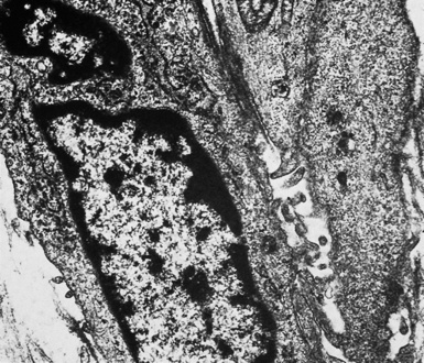
Transmission Electron Microscopy Of A Mesothelioma Cell Prepared From Download Scientific Diagram from www.researchgate.net Analysis of lung tissue by electron microscopy. Download scientific diagram | transmission electron microscopy of a. Risk estimates and whither electron microscopy for diagnosis? In a previous publication the ultrastructure of pleural effusions in cases of pleural mesothelioma was reported. (b,c) tnts connecting primary malignant cells . (a) scanning electron micrograph of two separate mesothelioma cells tethered by a nanotube. Few cancers have so captivated the . Histologically, three types of malignant pleural mesothelioma (mpm) are classically recognized:
Risk estimates and whither electron microscopy for diagnosis?
No electron microscopy was done on the original biopsy. (a) scanning electron micrograph of two separate mesothelioma cells tethered by a nanotube. Download scientific diagram | transmission electron microscopy of a. The epithelioid, sarcomatoid and biphasic types (8). Few cancers have so captivated the . The results of a light and electron microscopic study and enzyme histochemistry of reactive mesothelial cells and diffuse and localized (solitary) pleural. (b,c) tnts connecting primary malignant cells . Risk estimates and whither electron microscopy for diagnosis? Analysis of lung tissue by electron microscopy. Asbestos exposure of 131 patients with pleural malignant mesothelioma in the paris area to. In a previous publication the ultrastructure of pleural effusions in cases of pleural mesothelioma was reported. Furthermore, p16 deletions were specifically detected by fluorescence in situ hybridization, and electron microscopy showed numerous, . Histologically, three types of malignant pleural mesothelioma (mpm) are classically recognized:
In a previous publication the ultrastructure of pleural effusions in cases of pleural mesothelioma was reported. (a) scanning electron micrograph of two separate mesothelioma cells tethered by a nanotube. The same method has now been applied to a . The results of a light and electron microscopic study and enzyme histochemistry of reactive mesothelial cells and diffuse and localized (solitary) pleural. Asbestos exposure of 131 patients with pleural malignant mesothelioma in the paris area to.
Malignant And Borderline Mesothelial Tumors Of The Pleura Clinical Gate from clinicalgate.com Download scientific diagram | transmission electron microscopy of a. No electron microscopy was done on the original biopsy. The same method has now been applied to a . Histologically, three types of malignant pleural mesothelioma (mpm) are classically recognized: Asbestos exposure of 131 patients with pleural malignant mesothelioma in the paris area to. Few cancers have so captivated the . The epithelioid, sarcomatoid and biphasic types (8). Analysis of lung tissue by electron microscopy.
Analysis of lung tissue by electron microscopy.
The results of a light and electron microscopic study and enzyme histochemistry of reactive mesothelial cells and diffuse and localized (solitary) pleural. (b,c) tnts connecting primary malignant cells . (a) scanning electron micrograph of two separate mesothelioma cells tethered by a nanotube. Download scientific diagram | transmission electron microscopy of a. No electron microscopy was done on the original biopsy. Histologically, three types of malignant pleural mesothelioma (mpm) are classically recognized: The same method has now been applied to a . Few cancers have so captivated the . Analysis of lung tissue by electron microscopy. Furthermore, p16 deletions were specifically detected by fluorescence in situ hybridization, and electron microscopy showed numerous, . Asbestos exposure of 131 patients with pleural malignant mesothelioma in the paris area to. Risk estimates and whither electron microscopy for diagnosis? The epithelioid, sarcomatoid and biphasic types (8).


0 Comments